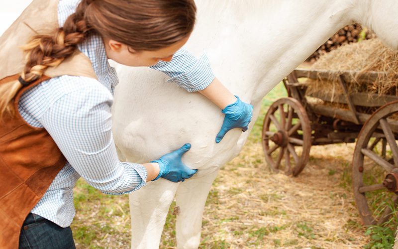
Pigeon fever (C. Pseudotuberculosis) and Ulcerative Lymphangitis in the Horse

What is Pigeon Fever?
‘Pigeon fever,’ also known as ‘dryland distemper’ is an abscess-causing bacterial disease of horses originally occurring in specific regions within California, Texas and Nevada. The bacteria, Corynebacterium pseudotuberculosis (C. pseudotuberculosis), has been confirmed in Utah, Colorado, Wyoming, Kentucky, Oregon, Idaho, Washington and Florida, as well as in Alberta, Canada.
The name ‘pigeon fever’ arises from the large swelling in the chest region, which resembles a pigeon’s breast. Pigeon fever is typically a disease of the late summer and fall months when conditions tend to be dry and dusty and populations of stable, horn and house flies peak. Recent reports suggest year-round occurrence of this disease in some regions.
How do horses get Pigeon Fever?
Bacteria gain entry into the horse through abrasions or wounds in the skin. Flies transmit the disease by carrying the bacteria from horse to horse and from contaminated soil to horse. C. pseudotuberculosis flourishes in manure-contaminated sandy and rocky soils, which serve as a major source of infection. Other means of transmission include direct contact with contaminated pieces of equipment, hands and contaminated hay or bedding. The incubation period, or time between exposure and appearance of clinical signs, is three to four weeks. The number of cases in a region fluctuates significantly from year to year, presumably due to herd immunity as well as environmental factors such as rainfall and temperature.
What are the clinical signs of Pigeon Fever?
In the western United States, pigeon fever infection results in external abscesses most commonly. External abscesses may grow as large as 8 inches (20 cm) prior to rupturing.
The most common sites and signs of external abscesses include:
- Swelling in the chest and bottom of the abdomen
- Hair loss and skin irritation around swellings, especially before abscess rupture
- Fever
- Lethargy/acting less energetic
- Decreased appetite
Less common abscess locations:
- Sheath and mammary glands
- Inguinal (between hind limb and body) and axillary (armpit) regions
- Smaller abscesses on trunk or limbs
Internal abscessation can also occur causing:
- Sites of internal abscess development in the lungs, liver, and/or abdominal lymph nodes
- Weight loss
- Organ-associated signs – colic, cough
An alternate form of pigeon fever called ulcerative lymphangitis affects the lymphatic vessels of the hind limbs. This condition appears to occur more often in Texas than in other states. In this painful form of the disease, multiple small abscesses and ulcers develop on the hind limb along the lymphatic vessels. One or both hind limbs may be affected, resulting in severe lameness, swelling and cellulitis (widespread infection of tissues under the skin), along with lethargy, fever and loss of appetite.
How is Pigeon Fever diagnosed?
History of either a single or multiple slowly-developing abscesses with the characteristic creamy whitish to greenish pus occurring during late summer to fall is supportive of a diagnosis of ‘pigeon fever.’
Veterinarians may obtain the purulent exudate (pus) from an external abscess or biopsies of affected skin or fluid retrieved from the airway or abdominal cavity of patients believed to have internal abscesses. Bacterial culture and polymerase chain reaction (PCR) of these fluid samples provide laboratory confirmation of pigeon fever.
Physical examination and bloodwork may be useful in making the diagnosis in some cases. The synergistic hemolysis inhibition (SHI) test evaluates protein titers in a patient’s blood and is particularly helpful in diagnosing the presence of internal abscesses in horses lacking external abscesses.
Ultrasound is useful to reveal suspected abscesses either in the lungs and abdomen or deep within the triceps or quadriceps muscles of severely lame horses.
How is Pigeon Fever treated?
Treatment of external abscesses: Treatment is individualized for each patient depending on severity of disease. Mature abscesses may be lanced by your veterinarian and flushed out to remove as much pus as possible. Abscesses deep in the muscle may require ultrasound-guided drain-placement to achieve drainage. All material drained and flushed from the abscesses must be collected and appropriately discarded, so that the area is not contaminated with more bacteria.
Use of antibiotics in treating external abscesses is only necessary when complicating factors are present, such as persistent fever, depression, lameness, or cellulitis. Use of antimicrobials in uncomplicated cases of external abscesses may prolong the course of disease. Use of hot packs, hydrotherapy (water therapy) or poultices may encourage maturation of external abscesses.
For internal abscesses and ulcerative lymphangitis: Antibiotics are required for patients with internal abscesses and ulcerative lymphangitis. Since the bacteria invade cells and are protected by both the pus and the thick capsule surrounding the abscess, treatment may be prolonged (weeks to many months) and should be directed by your veterinarian and/or consulting board-certified large animal internal medicine specialist.
Treatment of ulcerative lymphangitis should be immediate and aggressive, initially including a combination of antibiotics and anti-inflammatories. Physical therapy including the application of compression wraps, frequent cold-hosing, and hand walking are also beneficial. Antimicrobial treatment is commonly continued beyond resolution of abscesses and ulcers to reduce the incidence of relapse.
Following disease resolution or once abscesses have stopped draining, the majority of recovered patients will require no aftercare and pose no risk of disease transmission.
What is the prognosis for recovery?
- Of the three syndromes, prognosis for recovery is best (excellent) for patients with uncomplicated external abscesses.
- Internal abscesses carry a 30–40% risk of mortality as these cases are often challenging to diagnose. Early and accurate diagnosis is important for positive outcomes.
- In severe cases of ulcerative lymphangitis, the prognosis for return to full use is guarded due to recurrent swelling and cellulitis or secondary problems such as laminitis.
Edited by:
Freya Stein, DVM, DACVIM (LAIM)
April, 2020
Articles by Specialty
- Cardiology (19)
- Large Animal Internal Medicine (23)
- Neurology (17)
- Oncology (21)
- Small Animal Internal Medicine (29)
Articles by Animal
- Cats (35)
- Dogs (52)
- Farm Animals (5)
- Horses (12)
